100 Fascinating Biology Research Paper Topics for Students
Biology research is a serious analytical task that usually contains scientific findings, debatable questions, and detailed explanations. Students who are studying biology may get an assignment to find some interesting biological topics to do research for essays, term papers, and scientific reports.
Biology research is a serious analytical task that usually contains scientific findings, debatable questions, and detailed explanations. Students who are studying biology may get an assignment to find some interesting biological topics to do research for essays, term papers, and scientific reports.


It is quite a challenging and overwhelming task that takes pretty much time and effort. If your topic is not relevant, you won’t be able to include a scientific argument and proceed with further discussion. In this article, you will discover some topics for biology projects that will help you gain attention in a rapidly evolving field like this.
How to Choose Topic for Biology Research Paper?
Well, you’ve been researching for a while now, and you are ready to focus on a particular topic. Professors often ask students to write about something that has not been researched for a hundred times. Among all topics in biology, you should choose the one you are actually interested in. There are certain tips you need to follow before opting for your topic:
- Narrow down the subject matter. Before choosing an interesting biology topic for your research paper, you first need to identify a particular aspect of biology that interests you.
- Examine the existing research papers. You should conduct thorough research on the existing scholarly articles in order to gain a better idea about recent trends in the particular sphere of Biology.
- Brainstorm a particular area. By brainstorming ideas and thoughts, you may find the ideal research topic for biology to focus on.
- Conduct a preliminary research. By conducting preliminary research, you will check the amount of materials covering the selected topic. If you fail to find any information, you should choose another topic.
- Have a look at relevant examples. Checking credible examples is important for biology topics selection. This way, you will learn how to structure your research paper and go about the selected topic.
History of Biology
Do you consider covering the history of Biology in your research? Then, you may refer to the history of all life forms on the planet and explore how they have been researched over time. Here are some topics to dwell on:
- How archeology influences animal biology?
- The latest explanation of Darwin’s theory in modern science
- Edward Jenner and his contribution to the fight against epidemics
- The dead branches of evolution
- Exploring the importance of evolution factors
- Study of modern theories related to the origins of humankind
- The contribution of Antonie van Leeuwenhoek to the science of Biology
- The most significant milestones in the development of behavioral mechanisms in the late XIX – early XX centuries
- Can we trust the Natural Selection Theory? Does it work in the human world?
- The development of genetics over the last century
Immune System Biology Research Topics
The immune system, as our main defense against different diseases and infections, is one of the most important topics for discussion in Biology. Take a look at the following topic examples:
- The resistance capacity of the human immune system
- Why is vaccination important and what are its benefits?
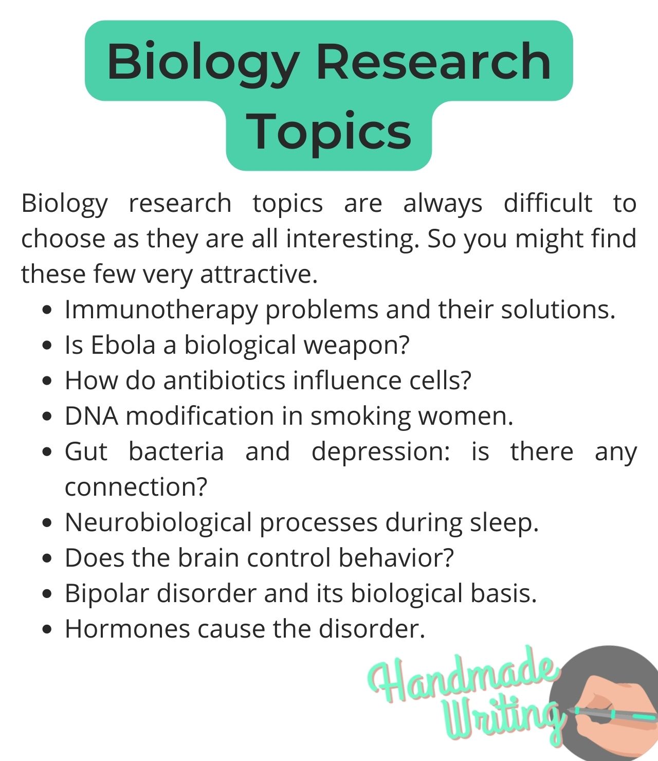
- Problems caused by immunotherapy
- Effects of probiotics on the prevention of infections
- How poor immunity leads to fatal diseases
- The functions of immune system agents
- The resistance of the human immune system
- Medical conditions caused by immune system malfunction
- How does insomnia influence the human immune system?
- Molecular biology of Human Immunodeficiency Virus
Molecular Biology Topics
Are you looking for a molecular biology issue to cover in your research paper? The following topics represent the latest research on this subject matter.
- The effective ways and tools for effective lifetime prolongation
- The role of genetically modified crops for the national economy
- Molecular biology research in the United States
- Can Ebola be viewed as a biological weapon?
- The effect on antibiotics on cells
- Challenges caused by diseases to molecular biology
- Does molecular biological research of rare genetic disorders provide us with the keys to understanding cancer and other diseases?
- What are the biological reasons behind food intolerances?
- Production of growth hormone and insulin in genetic engineering
- Molecular chaperones and their role in polypeptides folding
Genetic Researches Biology Topics
Research on genetic concepts can reveal intriguing insights into human nature and potential. The variety of options here is unlimited. Some of them include the following:
- How to solve the ethical dilemma of human cloning?
- The recent implementation of genetic disorders treatment
- Modern technology in DNA analysis
- The process of DNA modification on smoking females
- Methods of the sequencing of nucleotide sequences of DNA
- DNA Modifications and its Effects on Humans
- Genetics of chromosomal diseases related to structural chromosome rearrangements
- Genetics behind human physical features
- DNA diagnosis of hereditary and infectious diseases
- Can DNA influence the process of aging?
Neurobiology Research Topics
The human brain is intriguing, as there are always some new things to learn. The following topics have a great study potential:
- The improvement of brain activities with the most advanced neurobiology aids
- The innovative technologies in neurobiology
- Does gut bacteria lead to depression?
- Genetic defects that cause schizophrenia
- The molecular and gene regulatory signature of a neuron
- The influence of music on cognitive processes in a human brain
- What are the negative consequences associated with neurosurgery?
- Formation of thinking, speech, and consciousness of an average person
- Neurobiological Explanation of Sleep
- The role of neuroscience in the development of robotic technologies
Plant Research Topics
Are you interested in writing a paper about plants? Here are some of the latest ideas in botany to get inspiration by:
- How does climate change affect biodiversity in Australia?
- The evolutionary factors that affect plant growth
- A comparative analysis of invasive plants in New Zealand
- Friction in the plant world
- How modern technology can facilitate plant disease treatment
- Disease resistance mechanism in plants
- An extensive research on plant-associated microbes and available genomic tools
- Feature and functions of photosynthesis
- Impact of electric current on plant cells
- Plant cells plastids structure and function
Ecological Subjects
Ecology-concerning subjects are becoming more and more popular as society has to deal with the results of human behavior all the time. In your biology research study, you can offer some new solutions to ecological problems in order to turn the world into a better place. Let’s review some popular topic examples:
- Exploring the relevance of chemical ecology in the context of Oceania
- The impact of Ecological factors on animal behavior
- Ways animals and plants adapt to fast-changing environment
- Explore the ecological footprint of cotton production
- The ecological approach to sustainable marine research
- Why does biodiversity need to be conserved?
- Consequences of building the Hetch Hetchy valley dam
- What causes toxic algae bloom?
- Indoor air pollution: causes and risks
- The devastating impact of deforestation in Amazon forests
Zoology
If you are interested in the animal world , feel free to write about it in your research paper. By conducting a deep analysis of one phenomenon or species, you may shed light on the growing problems. Some of the burning topics to consider include the following:
- How the mechanism of camouflage is used by sea animals?
- How does veganism affect meat production?
- How do humans influence the diversity of animal species?
- The mechanism of resistance in animals
- Domestication: can foxes become domestic animals?
- Possibility of homosexual connections in the world of animals
- The future of the planet through the prism of Species Extinction
- Can beauty products testing on animals be viewed as ethical?
- Evolutionary connections between moths and butterflies
- The importance of home diet for dogs
Behavior and Hormones
You can also share ideas on how human hormones influence their mood and well-being. A short list of topic samples covers the following:
- How hormones affect women’s behavior during pregnancy
- Psychopathic Disorders: Are They Hormone-Specific?
- The hormones disorder and constant depression
- How does your brain control your behavior?
- The three main psychopathic disorders influenced by hormones
- Analyze the features of oxytocin that turn this hormone into a love drug
- How to generate growth hormone by means of genetic engineering methods?
- Biologic basis of the bipolar disorder
- The influence of diabetes and its potential threats
- The role of hormones in women’s health
Easy Biology Research Topics
If you don’t know what to write about in your biology research paper, you can use one of the most common topics. Although they have been widely covered by scientists, it doesn’t mean they are not suitable for further research. These topic ideas might come in handy:
- How to prevent the risks related to global warming?
- The future prospects of molecular biology in Europe
- Is growing organs for transplantation in laboratories ethical?
- How is melatonin used for therapy purposes?
- What is the value of sustainability in Biology?
- Explore the effects of marijuana on the human brain. Should the use of marijuana become legal?
- Accuracy of DNA testing in the modern medical environment
- Different ecological pyramids and the ratio of organisms at each of their levels
- The positive and negative aspects of transgenic crops
- Various means of wildlife protection
Conclusion
Biology research is one of the most complicated academic assignments that needs to be written according to strict requirements. While checking the variety of biology research topics, you should be ready to deal with certain problems, such as a poor understanding of the subject matter, its value for society, etc. Try to select a brief and concise topic that can be supported with relevant and up-to-date evidence. Make sure to conduct thorough research by using all the available tools and methods you know. Remember that the importance and timely revelation of the topic increases your chances to get an excellent grade eventually.
If you are still unsure on whether you can cope with your task – you are in the right place to get help. Our essay writers know the perfect answer to the question “Who can write my paper?”

A life lesson in Romeo and Juliet taught by death
Due to human nature, we draw conclusions only when life gives us a lesson since the experience of others is not so effective and powerful. Therefore, when analyzing and sorting out common problems we face, we may trace a parallel with well-known book characters or real historical figures. Moreover, we often compare our situations with […]
Read more...
Ethical Research Paper Topics
Writing a research paper on ethics is not an easy task, especially if you do not possess excellent writing skills and do not like to contemplate controversial questions. But an ethics course is obligatory in all higher education institutions, and students have to look for a way out and be creative. When you find an […]
Read more...
Art Research Paper Topics
Students obtaining degrees in fine art and art & design programs most commonly need to write a paper on art topics. However, this subject is becoming more popular in educational institutions for expanding students’ horizons. Thus, both groups of receivers of education: those who are into arts and those who only get acquainted with art […]
Read more...